Industry · August 21, 2026
Facial Nerve Injury After a Facelift: The Complication Every Surgeon Fears Most
A hematoma can be drained. A patch of dead skin heals, eventually. A facial nerve that has been cut is a different category of problem: the smile goes crooked, the brow stops lifting, and the patient has to live with it in the mirror every day. It is also rare, and most of what looks like it in the first weeks is not it at all. Here is how often facial nerve injury after a facelift really happens, which branches are at risk and why, what separates a bruised nerve from a divided one, and what a patient should hear from a surgeon before consenting to an operation that works within millimeters of the nerves that move the face.
By The Editorial Desk
11 min read
The facelift patient who wakes up and cannot raise one eyebrow has a long night ahead of her, and so does her surgeon. By morning she has tried to smile in the bathroom mirror and noticed that the left corner of her mouth lags. She has read, because everyone reads, that this can be permanent. The surgeon, for his part, is running back through the operation in his head, trying to remember every pass of the scissors near the zygomatic arch.
Most of the time, both of them will be fine. The weakness will resolve over days or weeks as a bruised nerve recovers. But not always, and the difference between those two outcomes is the subject of this article, the seventh in a series on how cosmetic operations go wrong, after the seroma, the hematoma, the incision that opens, the drain, fat necrosis, and skin necrosis. Facial nerve injury belongs at the end of the list because it is the one that cannot be fixed with a needle, a dressing, or patience. It is the complication that shapes how every facelift is designed.
What the facial nerve does and where it lives
The short answer: the facial nerve is the motor wire for every expression on your face, it exits the skull just in front of the ear and fans out across the cheek in five branches, and a facelift is performed in exactly the plane those branches run through.
The seventh cranial nerve leaves the skull at the stylomastoid foramen, behind and below the ear, and enters the parotid gland. Inside the gland it splits into its five named branches: temporal (or frontal), zygomatic, buccal, marginal mandibular, and cervical. Each exits the front edge of the parotid and travels forward under the SMAS, the fibromuscular sheet that facelift surgeons tighten, to reach the muscles it drives. The temporal branch lifts the brow. The zygomatic branch closes the eye. The buccal branch works the upper lip and the smile. The marginal mandibular branch pulls the lower lip down when you show your bottom teeth. The cervical branch runs the platysma in the neck.
This is what makes facelift surgery anatomically tense. The skin-only lift of the 1970s stayed above the SMAS and above the nerves, and rarely injured them; it also did not last. Every modern technique, from SMAS plication to the deep plane facelift, works in or under the SMAS because that is where the structural lift comes from. Working there means working on the same floor as the nerve. Surgeons learn a handful of landmarks in residency precisely to keep their instruments on the safe side of it: Pitanguy's line for the temporal branch, running from a point half a centimeter below the tragus to a point one and a half centimeters above the lateral brow; the zygomatic arch, where the temporal branch is protected by almost nothing; and the region two centimeters below the angle of the jaw, where the marginal mandibular branch dips before climbing to the chin.
How often it actually happens
The short answer: temporary facial weakness after a facelift shows up in somewhere between 1 and 5 percent of patients depending on technique and how hard anyone looks, and permanent injury is rare, with the large series clustering well under 1 percent.
The figure that anchors most consent discussions comes from a classic review by Baker and Conley of more than 6,500 facelifts, which reported permanent facial nerve injury in roughly 0.5 to 0.7 percent of cases, with transient weakness in a larger fraction. Later series have moved the permanent number down, not up. A 2020 systematic review in Aesthetic Surgery Journal pooled thousands of deep plane and extended SMAS cases and found temporary nerve weakness in the range of 1 to 2 percent and permanent injury in about 0.1 percent. A multi-surgeon review of extended deep plane facelifts published in Plastic and Reconstructive Surgery in the same period reported no permanent motor injuries across several hundred cases and transient weakness in about 2 percent.
Two qualifications are worth carrying around. The first is that these are surgeons who publish, which means they operate in volume and have usually been through their learning curve. Community rates are not tracked and are likely higher. The second is that "temporary" in the literature can mean anything up to a year. A patient whose smile is asymmetric for eight months will not be consoled by the fact that her case counts as a good outcome.
Which branches are injured is as predictable as which skin dies:
- The temporal branch is the most commonly injured in skin and SMAS facelifts, because over the zygomatic arch it lies just under the skin with only a thin layer of fat and fascia for cover. Injury shows as a brow that will not lift and a forehead that will not wrinkle on that side.
- The marginal mandibular branch is the most commonly injured in neck lifts and in the lower face, because it runs close to the jawline where platysma work and liposuction happen. Injury shows as a lower lip that does not pull down on one side, most visible when the patient shows her bottom teeth or says "eee." It is the injury most often produced by over-aggressive neck and platysma surgery.
- The buccal branches are injured more often than people assume, but they are multiple and cross-connected, so an injury to one is usually covered by its neighbors and goes unnoticed.
- The zygomatic branch is injured least often and matters most, because an eye that does not close is a medical problem, not a cosmetic one.
"Permanent facial nerve injury after a facelift runs well under 1 percent in published hands. Temporary weakness is several times more common, and the first two weeks cannot tell you which one you have.
"
Bruised nerve or cut nerve: telling them apart
The short answer: most early weakness is neurapraxia, a bruised or stretched nerve that recovers on its own within weeks to months, and the only way to know whether a nerve was actually divided is to wait, unless the surgeon saw it happen.
Nerve injuries are graded by depth. The mildest, neurapraxia, is a conduction block: the nerve is intact but stunned by stretch, pressure from swelling or a small hematoma, heat from cautery, or the local anesthetic itself. Function returns in hours to weeks as the insult resolves. The next grade, axonotmesis, means the axons inside the nerve have been damaged but the sheath around them is intact; the nerve regrows down its own tube at roughly a millimeter a day, so recovery takes months and is usually good. The worst, neurotmesis, is a divided nerve. Without repair it does not recover meaningfully, and the muscles it served slowly waste.
In practice this produces a rough clinical timeline. Weakness that appears in the recovery room and is gone by the first week was almost certainly anesthetic or swelling. Weakness that persists past two weeks but shows any flicker of movement is most likely neurapraxia and should keep improving. Weakness that is complete, with no movement at all, and has not changed by six to eight weeks is when a surgeon should be sending the patient for electromyography, which can distinguish a nerve that is conducting poorly from one that is not conducting at all. The American Academy of Facial Plastic and Reconstructive Surgery's guidance and most published algorithms agree on this point: observation for the first several months is appropriate, because the majority of injuries recover, and rushing to exploration in week three would mean operating on a great many nerves that were going to be fine.
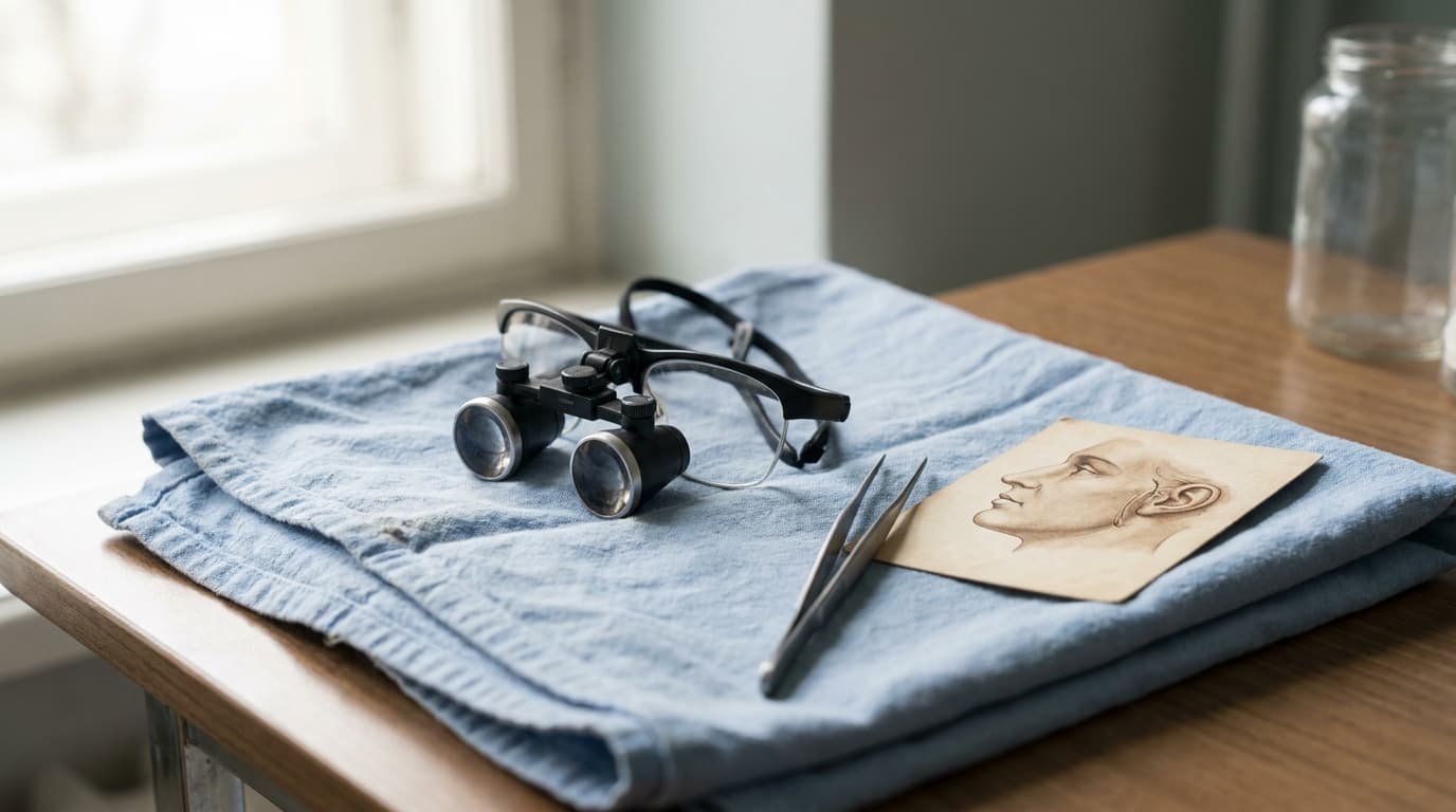
The exception is a nerve the surgeon knows was cut. A transection recognized during the operation should be repaired then and there, under magnification, because primary repair of a fresh, clean injury gives by far the best result. This is one of the quiet arguments for surgeons who operate with loupes or a microscope and who dissect under direct vision rather than blindly with scissors: you cannot repair what you did not see.
What the surgeon controls
The short answer: the dissection plane, the amount of traction, the use of cautery near known danger zones, and the discipline to stop when the anatomy is not what the textbook promised.
The paradox of modern facelift technique is that operating deeper, which sounds more dangerous, can be safer. In a deep plane or sub-SMAS dissection the surgeon lifts the SMAS as a sheet and works directly on top of the nerve branches, where they can be seen and protected. In a high-tension skin lift with blind SMAS plication sutures, the surgeon never sees the nerve and has to trust that a needle passed through the SMAS near the arch did not catch the temporal branch beneath it. Published series support the counterintuitive reading: the deep plane technique has not produced the nerve injury rates its critics predicted, and many of its proponents report lower transient weakness than in their earlier SMAS plication cases. The difference is direct vision.
Several specific habits separate careful surgeons from the rest:
- Staying superficial over the zygomatic arch. The temporal branch crosses the arch in the superficial temporal fascia, and a surgeon who keeps the dissection directly under the skin in that zone, or who stays deep to the fascia and lifts it as a protective layer, rarely injures it.
- Limiting cautery near the jawline and the arch. Thermal spread from electrocautery is a documented mechanism of nerve injury that leaves no visible cut. Bipolar cautery at low settings, or simple pressure, is the conservative choice near a branch.
- Respecting the retaining ligaments. The zygomatic and masseteric ligaments tether the SMAS, and nerve branches run close to them. Releasing them bluntly with a finger or a spreading motion, rather than cutting, is standard technique because it pushes a nerve aside instead of dividing it.
- Using the nerve stimulator selectively. Some surgeons map the temporal or marginal branch with a handheld stimulator before dissecting near it. It is not universal and is not a substitute for anatomy, but it costs little.
- Knowing the revision is harder. Secondary facelifts have scarred planes where the nerve can be stuck to the underside of the SMAS, which is one reason the revision consult deserves more scrutiny than the first.
Patient factors matter less here than in wound complications. Smoking does not predispose to nerve injury the way it does to skin necrosis. Prior facial surgery does, and so does prior parotid surgery or radiation, because the nerve has lost the fat that normally cushions it.
When it does not recover
The short answer: a weakness that has not improved by twelve months is treated as permanent, and there is a real, if imperfect, menu of options for restoring symmetry.
The first tools are not surgical. A brow that no longer lifts on one side can be matched by weakening the working side with neurotoxin, which lowers the good brow to meet the paralyzed one. The same approach works for the lower lip: a small dose of toxin into the functioning depressor on the opposite side evens the smile. It is a compromise, it needs repeating every few months, and it is what the majority of patients with a permanent single-branch injury end up choosing, because it is predictable. For the brow, a direct brow lift or an endoscopic brow lift on the weak side can restore resting position, though not movement.
Surgical reanimation is reserved for larger deficits. Nerve grafting or transfer, in which a branch from the unaffected side or another nerve is routed to the paralyzed muscle, can restore movement but is a major procedure with a year-long recovery. Static slings and muscle transfers, the operations developed for Bell's palsy and tumor surgery, are rarely needed after a facelift because the injuries are almost always partial. Facial plastic surgeons who specialize in facial paralysis, many of them affiliated with the AAFPRS and with academic centers, are the right referral, and a facelift surgeon who makes that referral promptly rather than reassuring a patient for a second year is behaving well.
The honest summary
Facial nerve injury is the facelift complication that nobody can patch, which is exactly why it is rare. The entire architecture of modern facelift technique, the landmarks, the planes, the blunt release of ligaments, the preference for seeing the nerve rather than guessing where it is, was built around avoiding it. In published hands, permanent injury is well under 1 percent, and temporary weakness, which resolves on its own in the large majority of cases, is in the low single digits.
The two numbers worth remembering are two weeks and twelve months. Weakness that is gone in two weeks was never a nerve injury in any meaningful sense. Weakness that is still there at twelve months is the real thing, and needs a plan. Everything in between is watching and waiting, which is the correct medicine and the worst possible experience.
Choose accordingly. Ask for a surgeon's own number, not the literature's. Ask which branch the planned technique puts at risk and how it will be protected. Ask what happens on the morning the eyebrow does not move. A surgeon who answers all three without flinching has told you more about the safety of the operation than any before-and-after photograph, because the photographs only ever show the smiles that worked.