Industry · August 9, 2026
The Specimen Nobody Mentions: What the Pathology Lab Finds in Tissue Removed During Cosmetic Surgery
A breast reduction, a tummy tuck, and an eyelid lift all remove real tissue from a real person, and somebody has to decide whether that tissue goes to a pathologist or into a bag. Published series find incidental cancer in a small but persistent fraction of breast reduction specimens. Almost no consultation covers who reads the report, who calls you, or what happens if it says something unexpected. Here is what actually gets examined, what gets discarded, and the one lab test that has to be requested by name.
By The Editorial Desk
8 min read
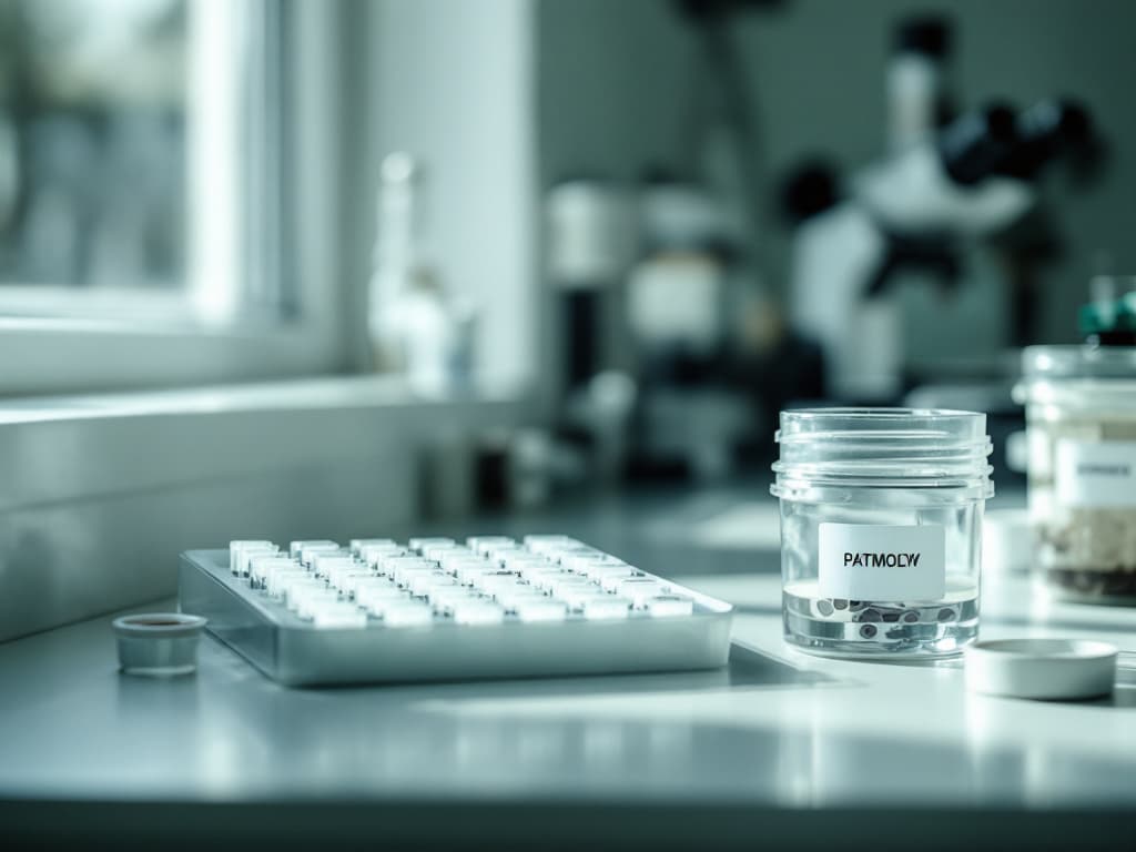
Cosmetic surgery removes tissue. That obvious fact has a consequence almost nobody discusses at the consultation desk: the pathology report after cosmetic surgery is a real document, generated by a real laboratory, that sometimes says something nobody was looking for. A breast reduction produces two specimens weighing several hundred grams each. An abdominoplasty produces a panniculus. A blepharoplasty produces strips of eyelid skin and small parcels of orbital fat. Someone in that operating room decides what happens to all of it, and the patient is almost never part of the decision or even aware one was made.
The number that makes this worth an article is small and stubborn. Published series of reduction mammaplasty specimens have reported incidental carcinoma or high-risk lesions in a range that runs from roughly one in a thousand to well over one in a hundred, depending on the population studied. Most large cosmetic-only series land under half a percent. The rate climbs sharply with patient age, and it is several times higher again in the symmetry reductions performed on the opposite breast of a woman already treated for cancer. Low, real, and entirely absent from the brochure.
Cosmetic operations generate specimens, and someone decides their fate
The short answer: some removed tissue is sent to a pathologist, some is examined only by the surgeon's eye, and some is discarded without either, and which happens depends on the procedure and the institution rather than on any single national rule.
The rough map looks like this. Breast reduction and gynecomastia excision produce tissue that is conventionally sent for microscopic examination. Abdominoplasty and panniculectomy specimens are often submitted, though the yield is low and some centers examine them grossly only. Blepharoplasty skin, facelift skin, and otoplasty cartilage are frequently discarded or examined without a microscope unless the surgeon saw something. Capsule removed during an implant exchange may or may not be submitted, and that turns out to matter more than the rest. Liposuction aspirate is essentially never evaluable at all, because the tissue arrives as an emulsified slurry with no architecture left for a pathologist to read.
None of this is scandalous. Hospitals and accredited facilities work from written lists, often modeled on College of American Pathologists guidance, specifying which specimen types may be exempt from routine microscopic examination because the diagnostic yield does not justify the cost. That is a legitimate resource decision made by people who have looked at the data. The problem is not that the lists exist. It is that the patient signing the consent form has no idea her operation involves a laboratory question at all, and therefore cannot ask the follow-up that actually matters.
"Every cosmetic operation that cuts tissue out is, incidentally and unintentionally, a screening event. Nobody markets it that way, and nobody should, but the patient deserves to know it happened.
"
The breast is where the incidental finding actually shows up
The short answer: reduction mammaplasty is the cosmetic operation with the highest documented rate of unexpected pathology, and age is the variable that drives it.
The published range is wide because the studies are not measuring the same populations. Series limited to young cosmetic patients report rates near zero. Series that include women in their fifties and sixties report figures around one to two percent for carcinoma or atypical proliferative lesions taken together. Contralateral symmetrizing reductions in breast cancer patients sit higher still, which is unsurprising given the underlying risk profile of that group. What survives across all of them is the direction: the older the patient, the more often the lab finds something.
This is why several groups have argued for a targeted rather than universal approach. Routinely submitting every specimen from every twenty-two-year-old is a poor use of pathology capacity. Reliably submitting specimens from patients over forty, and insisting on documented preoperative imaging in that group, is defensible on the evidence. The insurance and coverage mechanics that shape who gets a reduction in the first place are covered separately in the outcome data behind breast reduction surgery, and they interact with this: a threshold system that argues about grams removed rarely asks whether anyone looked at what was removed.
There is a quieter version of the same issue in male chest surgery. The excisional techniques described in what the evidence says about gynecomastia surgery produce a specimen. Pure liposuction reduction does not. Male breast cancer is rare, and the incidental yield in gynecomastia specimens is correspondingly low, but the structural point stands: choosing the technique also chooses whether a pathologist ever sees the tissue.
The capsule is a specimen with a special instruction attached
The short answer: when fluid or capsule is removed from around a breast implant, the test for implant-associated lymphoma has to be requested specifically, because routine culture and standard histology will not find it.
This is the sharpest practical example in the field. A patient with a late seroma around a textured implant needs the fluid sent for cytology with CD30 immunohistochemistry, and the capsule assessed accordingly. FDA communications and the NCCN consensus pathway both make that explicit. A surgeon who drains the fluid, sends it for ordinary culture, gets a negative result, and reassures the patient has performed a test that cannot detect the thing being ruled out. The broader risk picture, including which implant surfaces carry it and what the absolute numbers look like, is laid out in where the BIA-ALCL risk picture stands now.
The same logic runs through long-term implant care generally. The follow-up habits worth keeping are set out in what implant surveillance should actually look like, and the pathology question belongs in that list. Any late, unexplained swelling around an implant is a specimen waiting to be collected correctly, and the difference between correct and incorrect collection is a single line on a lab requisition.
On the face, the finding usually arrives before the specimen does
The short answer: eyelid and facial skin cancers are common, they present in exactly the areas cosmetic surgeons and injectors spend their time looking at, and the finding is more often made by the eye than by the lab.
Basal cell carcinoma is the most common malignancy of the eyelid, and the periocular region, nose, and temple are high-frequency sites for non-melanoma skin cancer generally. A surgeon planning a blepharoplasty is studying that anatomy under good light at close range for twenty minutes. Case reports and small series of incidental malignancy found in routinely submitted blepharoplasty and facelift skin exist, and the numbers are small, but the more useful version of this is not the lab result. It is the lesion the surgeon notices during planning and biopsies deliberately instead of excising blind as part of a cosmetic flap.
That cuts both ways, and the second edge matters more. A cosmetic consultation is not a skin examination, an injector is not a dermatologist, and nobody should be substituting a filler appointment for the annual check the AAD recommends for people with risk factors. The supervision problem described in who is actually injecting you applies here with force. A physician-led practice that spots something and refers is doing its job. A rotating-staff pop-up with no chart and no physician on site is not positioned to notice anything, and was never claiming to be. The pigment-related risks that make this harder to read in some patients are covered in where the risk actually sits for cosmetic procedures on deeper skin tones.
What happens after the report comes back is a systems question, not a clinical one
The short answer: the failure mode in pathology is almost never the lab getting it wrong, it is the result arriving somewhere nobody is watching.
Across medicine generally, failure to follow up on a completed test result is a well-documented category of harm and a recurring theme in malpractice claims. The mechanism is mundane. The report lands in a portal during a vacation, a surgeon assumes the office manager handled it, the office manager assumes the surgeon read it, and a patient who was told "we will call you if anything comes up" concludes correctly from the silence that nothing did.
Cosmetic practice has structural features that make this worse rather than better. Many aesthetic patients have no ongoing relationship with the practice after the final post-operative visit. Many have no primary care physician actively involved in the episode. Some travel for the procedure, which is one of the costs not itemized in the quote and one of the reasons plastic surgery tourism carries risk that does not appear on the invoice. And billing for pathology is frequently separate from the surgical fee, which is another line item that behaves the way the rest of the quote does: it exists, it is real, and it is not in the number you were given.
The fix is not clinical sophistication. It is a tracking process, a named person, and a default of telling the patient the result either way.
The honest summary
Tissue removed during cosmetic surgery is sometimes examined by a pathologist and sometimes is not, and the rules governing which is which are institutional, reasonable, and invisible to the patient. For most people this changes nothing, because most specimens are normal and most cosmetic patients are young enough that the yield approaches zero.
Three things are still worth carrying into a consultation. If your operation removes breast tissue and you are over forty, the odds of an unexpected finding are low but they are not theoretical, and both the pathology submission and your preoperative imaging should be settled questions rather than assumptions. If you are having anything drained or removed from around a breast implant, the lymphoma workup has to be named on the requisition or it does not happen. And whatever the specimen, the question that predicts your actual experience is not about the laboratory. It is about the office: who reads the report, and how you will be told.
A practice that answers that in one confident sentence has a system. A practice that has never been asked will tell you so with its face, and that is worth knowing before you sign anything.