Procedure Deep-Dive · September 8, 2026
Cosmetic Mole Removal: Shave Versus Excision, Why a Mole Can Grow Back Under a Flat Scar, and Why Every One of Them Belongs in a Pathology Jar Even When It Came Off for Looks
Removing a mole is the smallest operation in aesthetic medicine and the one most often done badly, because it looks too simple to plan. There are two honest ways to take a mole off: shave it flush with the skin and let the wound heal on its own, or cut it out whole and sew the line closed. Each leaves a different scar, each fails in a different way, and the choice between them is decided by the mole, the location, and the patient's skin far more than by preference. Underneath both sits a rule that the retail end of the market has started to skip: whatever comes off goes to a pathologist, because a mole removed for cosmetic reasons is still a pigmented lesion, and the only way to know what it was is to look. This piece covers what a mole actually is and which ones people want gone, what a shave does and why the pigment can return in a form that alarms pathologists, what an excision does and why the scar is three times longer than the mole, why lasers and pens and mail-order acids destroy the one thing that mattered, and where the scar will be worse than the spot it replaced.
By The Editorial Desk
21 min read
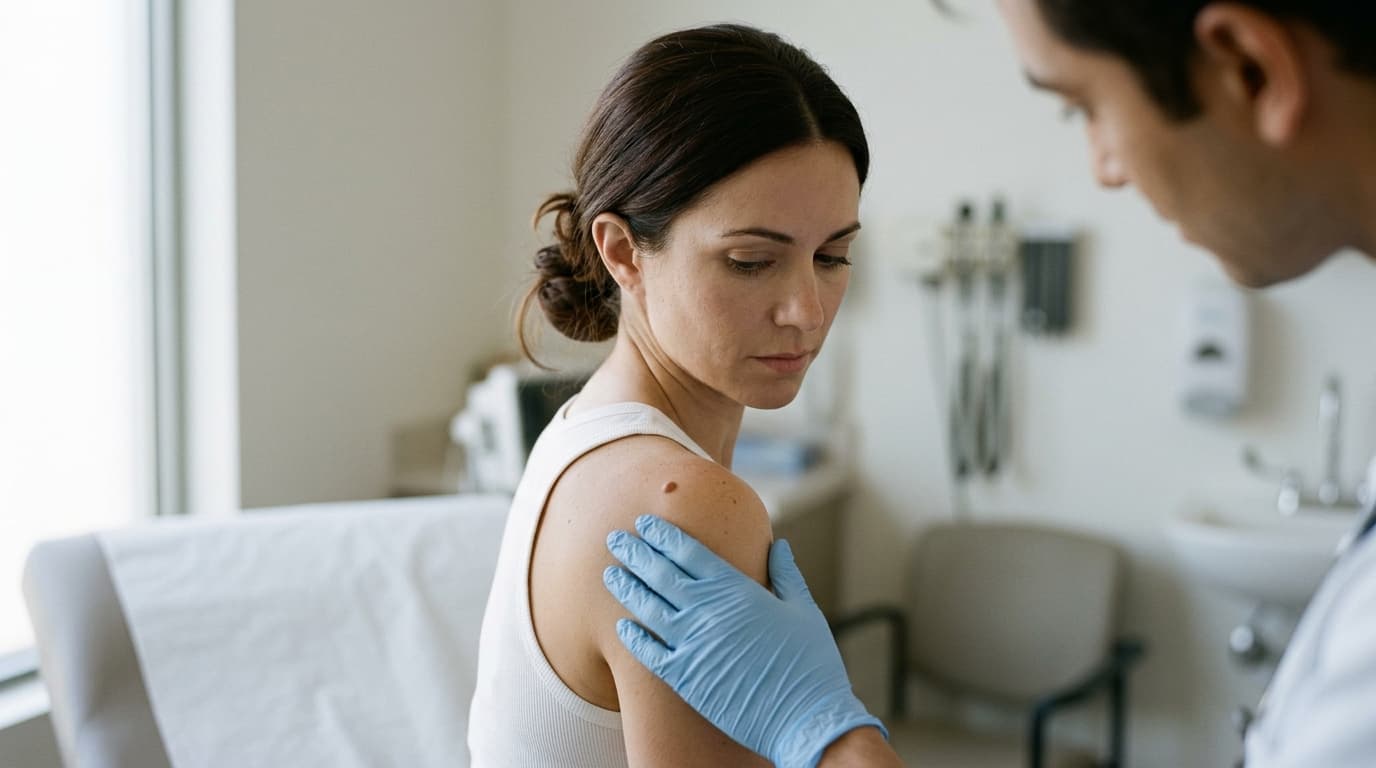
Ask a plastic surgeon or a dermatologic surgeon what the smallest procedure they perform is and most will say a mole. Ask them which small procedure generates the most unhappy follow-up visits and a surprising number will give the same answer. The mole that was supposed to vanish left a white coin on a tanned cheek. The mole on the shoulder came off cleanly and came back as a brown smudge inside the scar six months later. The mole on the chest was taken out with a neat ellipse and healed into a raised red rope three times as long as the spot it replaced. The mole that a spa burned off with a laser turned out, two years later and a few centimetres away, to have been something else entirely, and there was no specimen to check.
None of these outcomes is rare and none is mysterious. They follow from the fact that "mole removal" is not one procedure but two, that each is right for a different lesion in a different place on a different patient, and that the industry selling the service has an incentive to describe it as a quick tidy-up rather than as a small operation with a scar and a pathology report at the end. This piece is about the two honest methods, the dishonest ones, and how to tell which you are being offered. It sits alongside the piece on what a pathology report after cosmetic surgery actually says and the piece on reconstruction after facial skin cancer, which is where a small fraction of "cosmetic" moles end up.
What a mole is, and which ones people actually want removed
The short answer: a mole is a benign cluster of pigment cells (a melanocytic nevus) that sits at the junction of the epidermis and dermis when it is young and flat, and sinks into the dermis over decades until it is raised, soft, and often paler than it began; the moles people want removed for appearance are almost always the raised, mature, intradermal kind on the face, neck, and trunk, and a meaningful fraction of what patients call a mole is not a mole at all.
The average adult carries somewhere between ten and forty moles, most acquired in childhood and adolescence, and most of them spend their lives being ignored. The nevus starts as nests of melanocytes at the dermal-epidermal junction, which is what makes it flat and brown: the pigment is close to the surface. Over the years the nests migrate downward. A compound nevus has cells at the junction and in the dermis and is slightly raised. An intradermal nevus has abandoned the junction altogether, sits entirely in the dermis, and presents as a dome-shaped, skin-coloured or pinkish bump, often with a hair growing from it, on the cheek, the side of the nose, the neck, or the back. That is the mole that catches a razor, sits where glasses rest, or shows in every photograph, and it is the mole that walks into a cosmetic consultation.
This maturation matters for the surgical choice in a way patients are rarely told. A flat junctional nevus has its pigment near the surface and can, in principle, be removed by a shallow shave. A raised intradermal nevus has cells all the way down into the dermis, sometimes into the fat, and a shave that removes the visible bump can leave the roots in place. The depth of the mole is not visible from the outside. It can be estimated from its history and its feel, and it can only be confirmed by looking at the specimen under a microscope, which is the first of several reasons the specimen must exist.
Then there is the question of whether it is a mole at all. The lesions most often brought to a cosmetic consultation as moles include seborrheic keratoses, the waxy, stuck-on brown patches that accumulate on the trunk and face after forty and are not melanocytic at all; dermatofibromas, the firm brown buttons on the legs that dimple when pinched; skin tags, which are folds of skin on a stalk in the neck and armpits; and sebaceous hyperplasia, the small yellowish domes with a central dip that appear on oily foreheads and cheeks. Each is benign, each is removed differently, and none is a nevus. Also on the list, less often but more importantly, is basal cell carcinoma, which in its pigmented form can pass for a mole to a patient and occasionally to a clinician, and which the Mohs reconstruction piece describes in detail. A practitioner who cannot tell you, before removing your mole, which of these things they think it is has not examined it.
Shave removal: what it does, what it leaves, and why the mole can come back
The short answer: a shave excision numbs the skin with a small injection of local anaesthetic, takes the raised part of the mole off flush with or slightly below the surrounding surface using a flexible blade, seals the base with light cautery or a chemical styptic, and leaves a round wound that heals on its own in one to three weeks into a flat, usually pale or faintly pink circle about the size of the original mole; it involves no stitches and produces the least conspicuous scar on the face, and its known weakness is that it can leave nevus cells at the base, which regrow pigment in a proportion of patients and can do so in a pattern that mimics melanoma under the microscope.
The appeal of the shave is obvious once a patient has seen the alternative. There is no line, no suture marks, no incision three times longer than the mole. The wound is the footprint of the lesion and nothing more, and on a face it usually fades to something a stranger would not notice. The piece on numbing cream covers what a topical anaesthetic can and cannot do; for a shave the injection is the standard, it stings for a few seconds, and the procedure itself is over in a minute or two. Aftercare is petrolatum and a small dressing, and the piece on showering after cosmetic surgery applies in miniature: wet is fine, soaking is not, and the crust should be left alone.
What the shave cannot do is remove what it cannot see. The blade is set by the surgeon's eye at the level of the surrounding skin, sometimes a fraction below it. A junctional or thin compound nevus is gone at that depth. A mature intradermal nevus, with cells reaching well into the dermis, is decapitated rather than removed. Histological studies of shaved nevi have repeatedly found nevus cells present at the base of the specimen in a majority of intradermal lesions, and although most of those cells never do anything visible, some regrow pigment. The published rates of clinical recurrence range from a few percent to around a third, depending on the type of mole, how deep the shave went, and how long the patient was followed. Recurrence typically appears within the first six months as an irregular brown patch confined to the pale scar, and patients understandably assume something has gone wrong.
It is what happens next that gives this section its weight. A recurrent nevus, when biopsied, can show features under the microscope that resemble melanoma: irregular nests, pigment cells scattered up into the epidermis, and asymmetry, all confined to the scar. Pathologists have a name for this, pseudomelanoma, or the recurrent nevus phenomenon, described in the 1970s and now well recognised. The condition is benign, but the histology is alarming enough that the pathologist needs to know that the lesion was shaved before and what the original specimen showed. If the original shave was sent for pathology and read as a benign intradermal nevus, the recurrence is a nuisance. If the original was vaporised by a laser, burned off by a pen, or dissolved by an acid cream, and there is no earlier report, the pathologist is looking at melanoma-like cells in a scar with no history, and the patient may end up with a wide excision they never needed. That is the shave's real failure mode, and it is a failure of the specimen, not the blade.
The hair is the smaller disappointment. A shave removes the mole and leaves the follicle, so the hair that grew from a hairy nevus usually grows back from the scar. Electrolysis or laser hair removal can deal with it afterwards. And the colour is the third: a shaved site heals paler than the surrounding skin in most patients and can heal darker in some, particularly in the deeper skin tones the piece on cosmetic procedures on deeper skin tones covers, where the round white or brown coin is sometimes more visible than the mole was. A surgeon who has shaved a thousand moles will tell you which cheek, which skin, and which lesion will end up that way. One who has not will tell you it will disappear.
Excision: the ellipse, the three-to-one rule, and the line that replaces the spot
The short answer: a full-thickness excision removes the mole and a millimetre or two of normal skin around it as a spindle-shaped ellipse whose length is roughly three times its width, through the full depth of the skin into the fat, and closes the wound in layers with buried absorbable sutures and a fine surface stitch or skin glue; it removes the entire lesion with margins the pathologist can read, has the lowest recurrence rate of any method, and trades a round scar the size of the mole for a straight line about three times the mole's diameter, placed along the natural skin tension lines and healed as well as the location and the patient's skin allow.
The geometry is the part that surprises patients. If a six-millimetre mole is cut out as a circle and the edges are pulled together, the skin bunches at each end into the puckered cones the piece on dog ears after tummy tuck and liposuction describes at much larger scale. To avoid them, the surgeon draws an ellipse with pointed ends around the mole, and the mathematics of closing a curved wound flat require the ellipse to be about three times as long as it is wide. A six-millimetre mole therefore leaves a scar of roughly eighteen to twenty millimetres. On a cheek that line is oriented along the relaxed skin tension lines, which run roughly with the wrinkles a person will eventually have, so that it settles into a crease. On a back or a shoulder there is no crease to hide in, the skin is thick and under tension, and the line is what it is.
The closure is a small piece of proper surgery. Deep absorbable sutures take the tension off the skin edge and hold the dermis together while it heals; a fine running or interrupted surface suture, or an adhesive, approximates the edges. The surface stitches come out at five to seven days on the face and ten to fourteen days on the trunk and limbs. The deep sutures dissolve over weeks to months, and the piece on spitting sutures explains why one of them occasionally works its way to the surface as a small tender lump before it does. On a high-tension site like the upper back or the shoulder, an excision can also separate at the edges if the patient reaches or lifts too early, which is the miniature version of the problem the wound dehiscence piece covers. A surgeon who says "no gym for two weeks" after a mole excision on the deltoid is not being cautious for its own sake.
There is a middle path for small lesions. A punch excision uses a circular blade of a few millimetres' diameter to core out the mole through full thickness, and the resulting small round defect is closed with one or two stitches into a short line. It suits flat or slightly raised moles up to about the size of a pencil eraser in locations where an ellipse would be excessive, and it delivers a full-depth specimen to the pathologist. It also produces the small dog ears the ellipse was designed to avoid, so it is a compromise, not a trick.
The excision's advantage is complete in a way that the shave's is not. The entire lesion, top to bottom and side to side, is in the jar. The pathologist can report whether the margins are clear, and in the small proportion of moles that turn out to be atypical the question of whether more skin needs to come out is answered from the specimen rather than from a guess. Recurrence after a complete excision of a benign nevus is close to nil. What the patient buys with that certainty is a line, and whether the line is a better bargain than the spot is the subject of the section on scars.
Why every mole goes to pathology, even the one removed for looks, and why the pens, acids, and spa lasers drew an FDA warning
The short answer: dermatology and plastic surgery bodies, including the American Academy of Dermatology, hold that pigmented lesions should be diagnosed before they are destroyed and that tissue removed by shave or excision should be examined by a pathologist, because clinical inspection even by specialists misses a proportion of early melanomas, roughly a quarter to a third of melanomas arise in or beside a pre-existing mole, and the American Cancer Society estimates around one hundred thousand new invasive melanomas in the United States each year; a mole removed for cosmetic reasons has not been exempted from any of this by the patient's motive, and the lasers, plasma pens, and mail-order creams that skip the step, the last of which drew an FDA consumer warning and a round of warning letters in 2022, share the defining fault of destroying the only thing that could have said what the mole was.
The argument for skipping pathology is always the same: it looked benign, it had been there for years, the patient only wanted it gone, and the fee for the pathologist is an annoyance on a cosmetic bill. Each premise is partly true and the conclusion is still wrong. Looking benign is not a diagnosis. Studies of diagnostic accuracy have found that experienced dermatologists examining a lesion with the naked eye correctly identify melanoma somewhere around seven times in ten, and that a dermatoscope, the magnifying lens with polarised light that has become standard in dermatology and is still absent from many aesthetic clinics, improves that figure substantially but does not make it perfect. Having been there for years is true of most melanomas that arise within an existing mole. And wanting it gone is a reason to remove it, not a reason not to look at it.
"A mole removed for cosmetic reasons is still a pigmented lesion. The patient's motive changes the billing code. It does not change the cells.
"
The destructive methods are where the guidance is sharpest. Lasers, electrodesiccation, cryotherapy, radiofrequency, and the handheld plasma devices sold to spas all remove a mole by destroying it in place. There is no specimen. The pigment is gone, the cells may or may not be, and if the lesion was an early melanoma the evidence has been cooked. The dermatology position is that a melanocytic lesion should not be treated with a laser or any other destructive modality unless it has been diagnosed first, by biopsy if there is any doubt, and that the routine laser treatment of moles for cosmetic reasons is not appropriate. This is different from the laser treatment of sun spots the piece on lasers for pigmentation describes: a solar lentigo is a patch of excess pigment in normal cells, a nevus is a colony of abnormal ones, and a clinic that treats them with the same device on the same afternoon has collapsed a distinction the field spent decades building. There are, in the literature, case reports of melanoma diagnosed in or under a nevus that had been lasered years before, delayed precisely because the laser had removed the surface pigment that would have prompted attention. The piece on laser tattoo removal and scarring covers what the same devices do to skin when they are aimed correctly; aimed at a mole, the scarring is the lesser problem.
Pathology also decides what happens next when the news is neither clearly good nor clearly bad. A proportion of removed moles are read as dysplastic, or atypical, nevi: benign, but with architectural or cellular features that place them somewhere between an ordinary mole and a melanoma. Whether such a lesion needs a second, wider excision depends on the degree of atypia and whether the first removal reached clear margins, and a 2015 consensus of pigmented-lesion specialists concluded that mildly and, in many cases, moderately atypical nevi with clear margins need no further surgery, that mildly atypical nevi with involved margins can reasonably be watched, and that severely atypical lesions should be re-excised. A shaved mole that turns out to be moderately atypical with cells at the base is the commonest awkward result of a cosmetic removal, and the conversation about whether to go back for a wider excision is one the patient should have with someone who reads the report rather than the summary. The piece on the pathology report explains how to read one. The piece on medical records after cosmetic surgery explains why the patient should have a copy: the recurrent-nevus scenario above is solved by that piece of paper.
The cost of the pathologist's fee is real and should be stated up front rather than discovered on the invoice. It is a fraction of the cost of the removal and a smaller fraction of the cost of a wide excision for a melanoma that was missed. A practice that offers to waive it, or that does not mention it because its method produces nothing to send, has made a decision about your risk that was not theirs to make.
The methods that produce no specimen deserve their own accounting, because they are the ones being sold hardest. There is no over-the-counter product approved by the FDA for removing moles, skin tags, or seborrheic keratoses, and the mail-order market is older than the internet. Bloodroot pastes, sold as black salve or under gentler names, have been eating through skin lesions and the healthy skin around them since the nineteenth century, and the FDA has been warning about them for decades. What changed was distribution. Mole-removal creams appeared on the major marketplaces with thousands of reviews, and in August 2022 the FDA published a consumer update stating plainly that no over-the-counter product is approved for the removal of moles, seborrheic keratoses, or skin tags, that the products being sold could cause injuries and scarring, and that a mole treated at home could be a skin cancer left undiagnosed. The agency sent warning letters the same year to sellers including, notably, a large online marketplace, for marketing unapproved drugs for these purposes. The injuries it described were not exotic: full-thickness chemical burns, permanent pitted or raised scars far larger than the mole, and infections, on faces.
The pens are the newer version of the same problem. Marketed as plasma pens or fibroblast pens, and sold from a few dozen to a few hundred dollars, they generate a small electrical arc that vaporises tissue on contact. Used by a trained operator on the right indication they have a place in the energy-device landscape the skin-tightening piece surveys, with appropriate scepticism. Used on a mole, at home or in a spa, they do what a laser does with less control: the surface is charred, the pigment cells beneath are left to their own devices, the scar is a burn scar, and there is nothing to send to anyone. Patients who have used one on a mole and then developed a dark patch in the scar are in exactly the recurrent-nevus-without-a-history situation described earlier, and their next stop is a wide excision for a lesion that may have been benign all along.
The spa laser is the respectable face of the category and needs saying separately because it is offered by licensed businesses. A pigment-targeting laser aimed at a flat brown mole will lighten it, sometimes dramatically, and the patient leaves pleased. Whether the nevus cells are gone is unknown, whether the lesion was a nevus is unknown, and the lightened residue is harder to assess at the next skin check than the original was. A spa that offers it is not breaking a law; it is operating outside a standard of care that exists for the reasons set out above. The trademarked procedure names piece is relevant too: a "pigment correction treatment" with a brand name is still a laser pointed at a mole.
None of this means a mole must stay. It means the sequence is fixed. Someone trained to recognise skin cancer looks at it, with magnification, and says what they think it is. It comes off by a method that produces a specimen. The specimen goes to a pathologist. The report comes back and is kept. Then the scar is managed. Reverse any step and the procedure has become something other than mole removal.
Where the scar will be worse than the mole
The short answer: the face heals shave and excision scars better than anywhere else on the body because of its blood supply and its creases, while the chest, shoulders, upper back, and jawline are the sites most prone to thick, raised, hypertrophic and keloid scars, particularly in patients with deeper skin tones or a personal or family history of keloids; on those sites a small mole is frequently a better cosmetic outcome than the scar that would replace it, and a surgeon who says so is doing their job.
Scar quality is decided by three things the surgeon does not control and two they do. The three are location, skin type, and the patient's own scarring biology. Facial skin is thin, richly vascular, relatively slack, and full of natural lines; a well-placed excision on a cheek can be almost invisible at a year, and a shave on the nose or temple usually fades to a faint pale spot. The presternal chest, the deltoid, the upper back, and the angle of the jaw are the opposite: thick skin under constant tension from the movement of the shoulder and arm, and the anatomical sites where hypertrophic scars and keloids cluster. Keloids, which grow beyond the boundary of the original wound and keep growing, are many times more common in patients of African, Asian, and Hispanic ancestry than in patients of northern European descent, and they run in families. The piece on scar care after plastic surgery covers what silicone, pressure, and steroid injection can and cannot do afterwards; the honest position is that the time to think about a keloid on the chest is before the mole comes off, not after.
The two things the surgeon controls are technique and choice of method. On the face, a shave is usually the cosmetic answer for a raised intradermal nevus and an excision for a flat or suspicious one. On the trunk, the calculus inverts in an uncomfortable way: the shave leaves a round pale or dark coin that on a shoulder can look like exactly what it is, while the excision leaves a two-centimetre line in the skin most likely to make it thick. A surgeon who recommends leaving a benign mole on the chest alone is not being lazy. They have looked at the scar they would produce and judged it worse than the spot. The piece on how to read a before-and-after gallery applies here in miniature: ask to see trunk excisions at a year, not face shaves at six weeks.
Then there is the question of who is holding the blade. Mole removal is performed by dermatologists, plastic surgeons, facial plastic surgeons, and general practitioners, and increasingly offered by medical spas and aesthetic clinics whose staff may be trained in lasers and injectables and not in surgical excision or in the recognition of skin cancer. The piece on who is injecting you describes the supervision gap in the retail aesthetic market; the mole is the place where that gap can cost a diagnosis rather than a result. The board certification piece sets out which credentials mean what. The practical test is simpler than a credential: does the person about to remove your mole own a dermatoscope, use it, tell you what they think the lesion is, and send the specimen to a pathologist without being asked? If any of those four is missing, the rest of the consultation is beside the point.
The honest summary
A mole is a benign colony of pigment cells that starts flat and sinks into the dermis over the decades until it is a raised bump, and the raised bump is what people bring to a cosmetic consultation. A meaningful share of what patients call moles are seborrheic keratoses, dermatofibromas, skin tags, or, occasionally, basal cell carcinomas, and the person removing yours should be able to say which before they start.
There are two honest ways to take a mole off. A shave removes it flush with the skin, needs no stitches, and leaves a flat round mark the size of the mole that fades well on the face and less well on the trunk or in deeper skin tones; its weakness is that it can leave cells at the base that regrow pigment in a proportion of patients, sometimes in a pattern that mimics melanoma under the microscope and is only correctly read if the original specimen was examined. An excision removes the whole lesion as an ellipse three times as long as it is wide, closed in layers, with the lowest recurrence rate and readable margins, at the cost of a line that heals into a crease on the face and into a visible, sometimes thickened, scar on the chest, shoulders, and back.
Everything removed goes to a pathologist. Clinical inspection misses a proportion of early melanomas even in specialist hands, a quarter to a third of melanomas arise in or beside an existing mole, and the fee for the report is small next to the cost of the alternative. Lasers, plasma pens, electrodesiccation, and the creams the FDA warned about in 2022 all destroy the lesion in place and leave nothing to examine, which is why dermatology guidance says not to use them on moles at all.
On the face, the small procedure usually beats the mole. On the chest, the shoulders, and the upper back, particularly in a patient who forms thick scars, the mole frequently beats the procedure, and a surgeon who tells you so has earned the consultation fee. Ask what they think it is and whether it will be sent to pathology. Ask which method and what the scar looks like on that exact spot at one year. Ask what happens if the report is atypical. The clinic that answers all three has treated your mole as the small operation it is. The one that reaches for a laser has treated it as a blemish, and the one thing a mole is not is a blemish.